


Palabras clave
leucemia linfoblástica
linfoma
inmunohistoquímica
inmunotipificación
desórdenes linfoproliferativos en gatos
CD79a
CD3
Cómo citar
Resumen
Clínica Veterinaria: abordaje diagnóstico y terapéutico
ISSN: 2395-8766
Cómo citar este artículo:
- Pérez Enriquez JM, García Ortuño LE, Romero Romero L, Carrasco Reyes A, Aguayo Ávalos O. Uso de los marcadores CD79a y CD3 en la tipificación inmunológica de leucemia linfoblástica, secundaria al virus de leucemia viral felina. Clínica Veterinaria: abordaje diagnóstico y terapéutico. 2017;3(1).
Hallazgos clínicos. Al inicio del examen físico no presentó alteraciones significativas. Realizamos una prueba serológica para diagnóstico de SIDA y leucemia viral felina (FIV/FeLV combo test IDEXX), que resultó positiva a leucemia viral felina.
Tratamiento y evolución. Iniciamos un protocolo inmunomodulador y se establecieron revisiones constantes. El día 425 se nos reportó depresión, anorexia, linfadenomegalia generalizada y palidez en las mucosas orales. En el hemograma observamos leucocitosis (627.2 × 10⁹/L) por linfocitosis; 407.7 × 10⁹/L correspondían a linfocitos inmaduros (prolinfocitos y linfoblastos) lo cual asociamos a leucemia linfoblástica aguda, secundaria a leucemia viral felina. Debido al mal estado del paciente, los propietarios decidieron aplicar la eutanasia; remitimos el cuerpo al área de necropsias de la FMVZ-UNAM. Se detectó infiltrado linfoide en distintos órganos, así como una médula ósea hipercelular. En la inmunotipificación del corte histológico de médula ósea, encontramos positividad para CD79a, con lo que determinamos que la estirpe celular neoplásica corresponde a linfocitos tipo B.
Relevancia clínica. En los gatos, a diferencia de los perros, existe escasa información en la literatura con respecto a la sobrevida y el pronóstico de las neoplasias linfoproliferativas. La inmunotipificación es útil para complementar la estadificación histológica y contribuir con información para determinar el comportamiento de estos procesos neoplásicos, e incluso tiene un valor pronóstico importante.
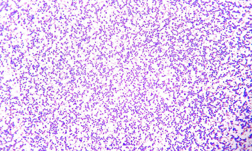
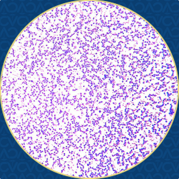
Figura 1. Frotis sanguíneo. Se aprecia una gran cantidad de leucocitos. 10x.Tinción de Wright
Use of CD79a and CD3 markers in the lymphoblastic leukemia immunological tipification secundary to viral feline leukemia virusCase description.A male European domestic cat of 5 months old and previously exposed to the FLV virus, was admitted into the Veterinary Hospital of the National Autonomous University of Mexico.Clinical findings. During physical examination, no significant alterations were found. A SNAP FIV/FeLV IDEXX Combo test was performed, which showed a positive result to FeLV.
Treatment and evolution. An immunomodulatory protocol was initiated and constant examinations were scheduled. On day 425, several symptoms such as depression, anorexia, generalized lymphadenomegaly and oral mucosa paleness were reported. A hemogram was performed, where leukocytosis (627.2x10⁹/L) was observed by lymphocytosis, from which 407.7x10⁹/L corresponded to immature lymphocytes (prolymphocytes and lymphoblasts) associated to acute lymphoblastic leukemia, secondary to viral feline leukemia. Due to the poor condition of the patient, euthanasia was performed and the body was referred to the pathology department of the FMVZ-UNAM for a post mortem study. Necropsy revealed lymphoid infiltrate in different organs, as well as, bone marrow hypercellularity. Bone marrow immunotyping was positive to CD79a, whereby it was diagnosed as a B-cell lymphoid neoplasia.
Clinical relevance. Unlike dogs, there is scarse information for cats regarding the survival and prognosis of lymphoproliferative malignancies. Therefore, immunotyping may be useful for complementing the histological staging and contributing with information, in order to determine the behaviour of these neoplastic processes, and even having an important prognostic value.
Keywords: viral feline leukemia, lymphoblastic leukemia, lymphoma, immunohistochemistry, immunotyping, lymphoproliferative disorders in cats, CD79a, CD3.
Referencias
Harvey WJ. 2012. Disorders of bone marrow. En: Veterinary Hematology a Diagnostic Guide and Color Atlas. St. Louis, Missouri, EUA: Elsevier. p. 298-–300.
Hartmann K. Feline leukemia virus infection. En: Green EC Infectious Diseases of the Dog and Cat. 4a ed. St. Louis, Missouri, USA: Elsevier; 2012. p. 113–8.
Vall DM, Young KM. Hematopoietic tumors. En: Withrow & MacEwen’s Small Animal Clinical Oncology. 4a. St. Louis, Missouri: Elsevier; 2007. p. 699–784.
Vail DM, Moore AS, Ogilvie GK, Volk LM. Feline lymphoma (145 cases): proliferation indices, cluster of differentiation 3 immunoreactivity, and their association with prognosis in 90 cats. J Vet Intern Med. septiembre de 1998;12:249–354. DOI: 10.1111/j.1939-1676.1998.tb02134.x.
Stansfeld AG, Diebold J, Noel H, Kapanci Y, Rilke F, Kelényi G, et al. Updated Kiel classification for lymphomas. The Lancet. 6 febrero 1988;331(8580):292–3. DOI: 10.1016/S0140-6736(88)90367-4.
Callanan JJ, Jones BA, Irvine J, Willett BJ, McCandish IA, Jarrett O. Histologic Classification and immunophenotype of lymphosarcomas in cats with naturally and experimentally acquired feline immunodeficiency virus infections. Vet Pathol. 1996;33:264–72.
Chino J, Fujino Y, Kobayashi T, Goto-Koshino Y, Ohno K, Nakayama H, et al. Cytomorphological and immunological classification of feline lymphomas: clinicopathological features of 76 cases. J Vet Med Sci. 21 enero 2013;75(6):701–7. DOI: 10.1292/jvms.12-0246.
Valli VE, Jacobs RM, Norris A, Couto CG, Morrison WB, McCaw D, et al. The histologic classification of 602 cases of feline lymphoproliferative disease using the National Cancer Institute working formulation. J Vet Diagn Invetigation. 1 julio 2000;12(4):295–306. DOI: 10.1177/104063870001200401.
Vezzali E, Parodi AL, Marcato PS, Bettini G. Histopathologic clasiffication of 171 cases of canine and feline non-Hodkin lymphoma according to the WHO. Vet Comp Oncol. Marzo 2010;8(1):38–49. DOI: 10.1111/j.1476-5829.2009.00201.x.
Petterson-Kane JC, Kugler BP, Francis K. The possible prognostic significance of immunophenotype in feline alimentary lymphoma: a pilot study. J Comp Pathol. 2004;130:220–2. DOI: 10.1016/j.jcpa.2003.09.008.
Comazzi S, Gelain ME. Use of flow cytometric immunophenotyping to refine the cytological diagnosis of canine lymphoma. Vet J. mayo de 2011;188(2):149–55. DOI: 10.1016/j.tvjl.2010.03.011.
Guzera M, Cian F, Leo C, Winnicka A, Archer J. The use of flow cytometry for Immunophenotyping lymhoproliferative disorders in cats: a restrospective study of 19 cases. Vet Comp Oncol. 2 junio 2014;14(S1):40–51. DOI: 10.1111/vco.12098.